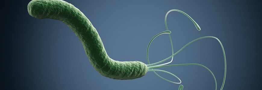
Helicobacter pylori
Peptic Predator
Stomach disrupter
Slithers under the surface.
Run silent, run deep.
Common knowledge. Conventional wisdom. Scientific consensus. Dominant ideology. Shared beliefs. Take it for granted. Some things in life, and medicine, are just accepted, probably because that’s how it’s always been, and no reasonable alternative exists. Some beliefs are held sacrosanct despite no compelling scientific evidence for support. Sometimes “facts” become so entrenched that their challenges are met with derision and ridicule.
It doesn’t happen often, but a commonly held belief is sometimes proven wrong. Achieving widespread acceptance of the new theory is frequently fraught with years of frustration, squabbling, and often acrimony. Fortunately, scientific evidence is the foundation for medical opinion, and eventually, the truth will prevail. Such was the case for a common human ailment, peptic ulcers.
Ulcers were once common. They were mainly life-inhibiting, with bouts of intense stomach pain that could last several days. Often there was nausea, bloating, and loss of appetite. These episodes would come periodically and were usually treated with antacids and a “bland” diet.
Some ulcers were more serious. They were marked by intense, unremitting pain and hemorrhage. Surgery was sometimes necessary to stop the bleeding. A peptic ulcer could be fatal if medical intervention was unavailable or unsuccessful. Rarely, a patient with a peptic ulcer developed stomach cancer. Many suffered.
Such a common, painful, and often severe medical condition required an explanation. For many years the one widely accepted was the production of excess acid in the stomach. Stomach fluids are acidic under normal conditions, but it was supposed that in some people the contents were more acidic than typical. This excess acid was held responsible for the erosion of the stomach and duodenum lining, leading to ulcer formation. The reason for the extra acid formation was often attributed to the nervous state of the patient: some people, because of the stress in their life, produce more stomach acid and, as a result, suffer more from ulcers. The common term “case of nerves” was often used.
Proving such a hypothesis is obviously tricky, but some tried. In one experiment, mice were outfitted in little mouse straitjackets and lowered into near-freezing water, clearly a stressful situation. Their stomach contents were then measured for acidity, and, sure enough, it could be concluded that stress resulted in the formation of excess stomach acid.
With such a straightforward explanation for the malady, the remedy seemed clear. Consume antacids, cut down on stress, and eat bland, non-spicey foods to reduce the acidity of the stomach contents. The stress reduction part was the least medically controllable. It was not uncommon for ulcer sufferers to have their stress levels enhanced by thoughtless people essentially blaming them for their own trouble. “Calm down, you’ll aggravate your ulcer!” “Your ulcer’s all in your head!” Needless to say, this didn’t help.
Barry Marshall was the ultimate “free-range” kid. He was born in 1951 in Kalgoorlie, a gold mining town 400 miles east of Perth, Australia. His father worked as a mechanic, and his mother was a nurse. They often moved around from job to job, and little Barry was never at a loss for adventure. He was a clever boy and had an insatiable need to find out how things worked. His father often accommodated him, showing Barry the inner workings of the machinery he worked on. Young Barry also had a great interest in his mother's medical books. After graduating from college, he chose to enter medical school. Internal medicine with an interest in research was his career goal.
Dr. Marshall spent the third year of his medical rotation at Perth Hospital in gastroenterology. Third-year doctors were required to do an investigative study, and around that time he met Dr. Robin Warren, a pathologist. The two struck up a friendship and often met in Dr. Warren’s office in the hospital basement. During their conversations, Dr. Warren mentioned that he frequently observed some unusual, curved bacteria on biopsy specimens of stomach tissue submitted because of possible stomach cancer. At the time, the stomach was believed to be free of infection by bacteria because of the level of acidity present and the inhospitable environment it presented. By the early 1980s, with a hundred years of investigation into the causes of infectious diseases, no one had proposed that bacteria could cause a stomach infection. Coincidentally, a few years before, another small, curved bacteria, Campylobacter, had been proven to cause gastroenteritis, a disease similar to that caused by Salmonella. No one had seen that one coming either. Dr. Marshall was intrigued and got a list of patients with the mysterious stomach bacteria from Dr. Warren.
After doing a good deal of investigating, Dr. Marshall concluded that there likely was an association between these curved stomach bacteria and peptic ulcers. But there was trouble in proving it. The first step in demonstrating an organism’s association with disease is the isolation and characterization of the bug. For bacteria, this usually means growing it in pure culture (the term “pure” means by itself, independent of other organisms). In 1982, the mysterious organism was only seen under the microscope in a pathology lab, so more work was needed. Dr. Marshall worked with members of the microbiology lab to try to grow the organism like other pathogenic bacteria they commonly worked with. Clinical specimens from biopsies and gastric washings were inoculated to solid culture media and placed in an incubator. After one and two-day incubations, the cultures were examined for bacterial growth. A simple, routine bacterial culture, but since the organism had never been described, it was hit-and-miss. The first attempts were unproductive.
Sometimes the culture plates were overgrown by non-pathogenic bacteria. Common bacteria can’t survive long or multiply in stomach acid, but they can survive transiently. Bacteria on recently consumed food or resident flora of the mouth that is swallowed can easily gum up a culture of stomach material by growing rapidly and abundantly, overwhelming the culture media. Typically, culture plates are held in the incubator for 48 hours, and if no growth on the plates is observed, the cultures are discarded with the report “no growth in 48 hours.” Such was the case in the first attempts to isolate the observed but not cultured stomach bacteria: overgrowth by commensal bacterial flora or no growth at all.
Sometimes, luck, or fate, intervenes. One day some cultures of stomach biopsy material were placed in the incubator as usual. But it was just before the Easter holiday, and the technician responsible for examining them took a few days off, and the culture plates were left unattended for several days. Lo and behold, when the plates were examined after an extended incubation, there were these mysterious-looking colonies. The same curved rod-shaped bacteria observed in the biopsy specimens were seen under the microscope when smears were made from the colonies. A prolonged incubation period was a necessary but unknown requirement for growing the organism.
We now know that the organism, Helicobacter pylori, has some demanding growth requirements in the laboratory, what clinical microbiologists call “fastidious.” In the lab, they must be grown on nutrient-rich culture media supplemented with whole animal blood. They require oxygen, but it must be in reduced tension, only about 2-5% as opposed to the nearly 20% of room air (the requirement for reduced amounts of oxygen is called microaerophilic), and they need elevated levels of CO2 (capnophilic). Also, on primary isolation from stomach material, the colonies take 5-7 days to grow, and even then, the colonies are small. They are translucent in appearance.
Now that the organism had been retrieved and described in laboratory culture, the next step was demonstrating that it could cause gastritis and peptic ulcers. It sounds simple enough, but there is one fundamental problem: common laboratory animals like mice and rats aren’t infected by the organism. Dr. Marshall had treated several patients diagnosed with peptic ulcers with a regimen of antibiotics, not only getting them through an ulcer episode but also curing their ailment. With such promising results and no susceptible laboratory animals, the next move seemed obvious: give the organism to an uninfected human, see if the disease occurred, then try to cure it with antibiotics. But medical ethics being what they are, only one person could be infected: himself.
There was one slight hitch. Dr. Marshall was married, and he and his wife had four children. His wife, Adrienne, was a highly literate scientist and knew her husband’s work well. In mulling it over, Dr. Marshall knew he would have to make a request of his wife. After pondering it for a while, he figured asking her forgiveness would be more fruitful than seeking her permission. So, after a biopsy showing he was organism free, it was down the hatch.
It didn’t take long for his question to be answered. In a few days, he was overwhelmed by severe stomach pain, nausea, and vomiting. He looked and felt like death warmed over. There was no hiding this from his wife, so he fessed up. In addition to Dr. Marshall’s health, her concern was for that of her children and their friends and acquaintances—this journey into the arena of scientific experimentation needed to cease. After a biopsy showed the presence of the organisms in his stomach tissue, Dr. Marshall took a regimen of antibiotics and was bug and symptoms free in a few days. His findings were revolutionary.
News of an event like that travels fast. At five o’clock one morning, an American writer called Dr. Warren, the pathologist, and asked him about the experiment. Dr. Warren, given to some hyperbole, told him it was true, adding that Dr. Marshall nearly died. It turned out the writer was not from a major reputable newspaper; he wrote for the “Star,” a popular American tabloid usually seen in supermarket checkout stands and given to stories about such things as alien babies. But the word was out about the cause and cure of peptic ulcers. The trouble was that it wasn’t accepted within the medical community.
Major breakthroughs in the field of medicine are rare, and when they occur, the news of their discovery follows a pattern. Major research centers using collaborative efforts by leading authorities issue press releases, hold news conferences, and experts not affiliated with the studies are interviewed and offer their support. In discovering Helicobacter pylori as the primary cause of gastritis, peptic ulcers, and stomach cancer, the leading investigators were two previously unknown individuals from Perth, Australia, with the worldwide announcement of the research first appearing in The Star supermarket tabloid. To say there was reluctance to accept the conclusions of the research is a tremendous understatement. Drs. Marshall and Warren had great trouble publishing their work in professional peer-reviewed journals and were often denied speaking engagements at international gastroenterology conferences. There reportedly were times when Dr. Marshall got up to speak, and attendees made a show of walking out of the venue. Radically changing accepted dogma is a challenging task.
In Australia, the acceptance of the work was easier, especially when the clinical success was so overwhelming. Eventually, researchers worldwide confirmed the nascent work done in Australia. It is now universally accepted that Helicobacter pylori is a prominent inhabitant of the human stomach worldwide, often causing gastritis, peptic ulcers, and occasionally stomach cancer. For their epic discovery, Drs. Barry Marshall and Robin Warren were awarded the Nobel Prize for Physiology and Medicine in 2005.
Helicobacter pylori is a crafty little creature. There is good evidence that it once colonized most humans, often causing debilitating, sometimes fatal, disease. Bacteria were first observed as causes of human illness in the 1880s, but H. pylori wasn’t described as a potential pathogen until 1982. So, for 100 years, it was hiding in plain sight. The primary reason for the oversight is the organism’s habitat within the body, the stomach and the upper small intestine (duodenum). These organs are awash in hydrochloric acid and digestive enzymes. Acid effectively kills bacteria, so to have an organism not just surviving but flourishing in this area seemed preposterous. But the bug is equipped with unique factors that make this remarkable feat possible.
Neutralizing the acid within the stomach is the top priority for Helicobacter. One of the breakdown products of protein is urea, a carbon with two amino groups attached. Helicobacter has a powerful enzyme, urease, that takes apart urea, yielding ammonia and carbon dioxide. The ammonia produced neutralizes acid, much to the benefit of the organism. The bug can also degrade short peptides with the same effect.
The physical features of Helicobacter give it an advantage in its environment. It has a spiral shape, allowing it to slither into nooks and crannies. It also has three powerful flagella, propelling it at great speed and enabling the bug to escape engulfment by neutrophils.
At some point, though, the organism must attach itself to cells lining the stomach. To do this, the organism has over a half dozen adhesins that allow the bug to attach itself tightly to the cells lining the stomach, the extracellular matrix surrounding these cells, and the layer of mucin covering the stomach lining. By attaching to these surfaces, the toxic materials of the bacterium can be more easily transferred into the infected cells. In this infusion of toxins into host cells, the greatest damage of the organism to the stomach lining is seen.
Helicobacter has several toxins that can damage human cells, but two predominate. One is called cytotoxin-associated gene A, or CagA. The other is vacuolating cytotoxin A or VacA. CagA and VacA make a formidable one-two punch when both are present.
Helicobacter contains a secretion system used to inject toxin into a cell lining the stomach or duodenum. It acts similarly to the one found in Salmonella: the organism attaches to the human cell, a spike is formed outside the bacterium, the spike penetrates the host cell, and the toxin is injected into the cell. CagA is introduced into gastric and duodenal epithelial cells in this way. VacA attaches itself to the outside of the host cell, then enters. Once inside the human cells, the toxins begin their mischief.
CagA binds to and disrupts the actin cytoskeleton of cells. It also interferes with the junctions between cells. The affected cells round up and lose their form. Signaling inside the cell is altered, and the cell is greatly discomposed. VacA creates channels in the host cell membrane, so material from inside the cell flows out. It also can initiate apoptosis in the affected cell.
The net result of the activity of the Helicobacter toxins is the release of substances from the cell that the organism needs to survive. An important one is urea, which the bacterium can metabolize to neutralize stomach acid. Another is iron, an essential element for microbial growth.
The inflammation or erosion of the cells lining the stomach and duodenum is a condition known as gastritis. When Helicobacter invades and colonizes the stomach, gastritis results. The symptoms experienced are highly variable. Some individuals, like Dr. Marshall, become violently ill. Others have mild discomfort, and many have little or no symptoms. Some afflicted patients develop peptic ulcers, and a small percentage will develop stomach cancer. Once Helicobacter colonizes the stomach and settles in, it’ll be there for life. The immune system is not able to clear it. Thankfully, though, antimicrobial agents that do a very good job of eliminating the pathogen are available.
There is clearly a wide display of symptoms in people whom Helicobacter pylori has infected. The reason why some are barely ill while others are severely affected can be summed up in two words…it depends. Helicobacter is extremely variable in its genetic makeup. In fact, just like fingerprints and snowflakes, no two strains infecting people are alike. Even in members of the same family who presumably infected each other, there are subtle differences in the genetic constitution of each person’s bacterial strain. The organism’s genome is subject to copying errors. Some people have strains with CagA and VacA toxins, some with only one, and others with neither. The molecular arrangement of the toxins can also vary, as can the attachment molecules' structure and the urease enzyme's strength.
People vary as well. Some have more acidic stomach fluid depending on their genetic makeup and diet. Our immune systems also show variability, with the power of cytokines differing among people. Neutrophils have a significant role to play in ulcer formation. Neutrophils are attracted by interleukin 8. In some people, the attraction of IL-8 is much stronger than in others, so the number and strength of neutrophils vary from one person to another.
Several organisms cause the illness known as gastroenteritis. The most common are members of the genera Salmonella, Shigella, Campylobacter, and select types of E. coli. The infections caused by these differing organisms follow a similar pattern: The organism is ingested, attaches to a segment of the bowel, unleashes its pathogenic brew of virulent materials, and the infected person’s immune system counteracts the invader. Typically, after several days or a week of illness, the pathogen is removed, and the patient returns to health. Occasionally the individual becomes a carrier of the organism, shedding it with no ill effects to themselves. But for the most part, the infection is soon over, and the bug is gone.
With infection by Helicobacter, things are different. Soon after it infects, the patient experiences gastritis. The severity varies. It may present as a mild “upset tummy” or be a severe case of nausea, vomiting, bloating, and loss of appetite. As in cases of gastroenteritis caused by other organisms, the immune system is alerted, and a challenge to the invading microbe is mounted. The difference with infection by Helicobacter is that the organism is not cleared. The organism repels the immune response, and it stays in residence in the stomach. With other GI infections in which organisms not eliminated achieve a carrier state, the infecting bug rarely causes reinfection. Without laboratory tests, the patient is unaware that they still are colonized by the bacteria. But Helicobacter, once established, usually causes recurrent symptoms for many years.
Helicobacter invades the cells and mucin layer lining the stomach. With its flagella and spiral form, it slithers through the mucosal layer at considerable speed, making it a difficult target for neutrophils and macrophages. When it adheres to the epithelial cells it is firmly attached, which also impedes the pursuing phagocytes. To clear it, the adaptive immune system must be engaged and vigorous. Most pathogenic organisms can thus be readily cleared, but through the eons, Helicobacter has adapted adroitly to its encounter with the cells and molecules designed to eliminate it.
Lymphocytes known as CD-4 T-helper cells play a big role in controlling infections. Among this group of lymphocytes, the Th1 group predominates in the cause to eliminate Helicobacter. When active and engaged, the TH1lymphocytes release interferon-gamma, a powerful attractant and activator of macrophages. Ideally, the macrophages will swoop into the area and engulf the invading bacteria, helping to end the infection. TH2 lymphocytes also are involved, inducing B-lymphocytes to produce antibodies. Another set of CD4 T-helper lymphocytes, known as the TH17 group, serves to attract neutrophils, which add to the elimination of the organism. Of course, the toxicity of the macrophages and neutrophils is high, and the process must be strictly regulated. The CD4 lymphocytes carrying out this job are the regulatory T-cells, the Treg cells. They can dampen the inflammatory process and ultimately help prevent tissue damage by macrophage and neutrophil “friendly fire.”
Helicobacter notably disrupts the activity of the Th1 lymphocytes. The crux of the lymphocytes’ activity is its ability to detect the invading bacteria and then send chemical signals to other immune system cells, such as macrophages. That’s why they are called “helper” cells. The bacterial toxin VacA interferes with this process by inhibiting the chemical signals sent to the lymphocyte’s nucleus to unleash the genes responsible for cytokine and interferon production. The net effect of this VacA interference is a diminished number of substances, such as interferon-gamma, produced. Without that attraction signal, the number of macrophages entering the infected area is reduced, as is the overall immune response.
Once activated by encountering the microbial antigen they are programmed to detect, lymphocytes normally begin to reproduce, making many more copies of cytokine-producing cells directed at the invading organism. Helicobacter produces an enzyme, GGT, that arrests the proliferation of lymphocytes. A second enzyme, arginase, metabolizes the amino acid arginine. For the bacterium, arginase accomplishes two tasks. Arginine is necessary for lymphocyte reproduction, so when it is in short supply, the cells don’t replicate as efficiently as they should. In addition, when the bacterial enzyme metabolizes arginine, ammonia is produced, assisting the bug in counteracting the amount of acid in its environment.
Not only are the lymphocytes inhibited in their quest to send chemical signals and to reproduce, but Helicobacteralso induces their death by apoptosis.
The result of these bacterial actions directed against the T-helper Th1 cells is a standoff. The lymphocytes keep coming and are not entirely destroyed, but their activity is much less robust than needed. Helicobacter is able to persist.
Neutrophils, in addition to macrophages, play a major role in ridding us of invading bacteria. The eradication of Helicobacter from our stomach lining is no exception. To be involved, neutrophils must be attracted to the area, and a major factor in their migration is the activity of another CD4 lymphocyte, the Th17 cells. They’re called Th17 cells because, when activated, they release the cytokine interleukin-17 (IL17), a powerful attractant for neutrophils. When lots of neutrophils swarm into an infected area, they gobble up the invading microbe, helping end the infection. However, Helicobacter has another trick it employs to help ensure its survival. It produces a molecule, IDO, that down-regulates the production of IL-17 by Th17 cells. That means fewer neutrophils than needed enter the infected area, giving the bacterium the advantage.
The T-helper lymphocytes must be tightly regulated lest they become overreactive and initiate tissue damage. Another T-helper CD4 lymphocyte carries out the control of the group, the one called the regulatory T-cell, usually abbreviated Treg. The Treg cells send down-regulating chemical signals to active CD4 lymphocytes, such as the Th1, Th2, and Th17 varieties, slowing down their activity. Without this control, there could be an overabundance of an immune response leading to tissue damage. Helicobacter has achieved the ability to encourage the conversion of naïve T-cells (those not yet directed to become a specific type of cell) to become Treg cells. This overabundance of regulatory cells further dampens the immune response, favoring the bacterium.
Helicobacter pylori has been an uninvited companion of humans for thousands of years. An unusual bacterial predator, it doesn’t cause an overwhelming, life-threatening disease in most people. In many, it persists unnoticed for the life of the infected person. Just how serious the infection can become depends upon several factors, including the organism's and the infected person's genetics. Worldwide, most individuals become infected as children, and their immune responses differ subtly from adults. As the infected child matures, the bacterium adapts until a balanced, steady-state existence is formed between the organism and host. Situations that disrupt that balance can lead to the emergence of significant diseases, including peptic ulcers and cancer. Stress is one factor that can diminish the immune response's activity, so perhaps the adage that high stress exacerbates peptic ulcers was on the mark.
Helicobacter is a carcinogen. Indeed, not everyone infected by it develops cancer, but some do. Two types of cancer predominate, adenocarcinoma and lymphoma. Besides the presence of the correct strain of the organism, many factors contribute, such as salt in the diet, genetic predisposition, other bacteria colonizing the stomach once Helicobacter raises the pH, and the amount of ulceration induced by the organism's activity and the immune system’s response.
With the disruptive and long-term effects of the organism on the components of the immune system, there is reason to believe that Helicobacter infection can be associated with auto-immune diseases. Such associations are difficult to prove, and the mere presence of the organism in an afflicted patient is not proof of cause. But studies are ongoing.
Like most bacteria, Helicobacter can be treated relatively easily with antibiotics and antimicrobial substances. The Centers for Disease Control in the U.S. recommends that infections without symptoms do not need to be treated. In cases that require therapy, CDC suggests a multi-drug regimen. Reducing stomach acid with a proton-pump inhibitor (such as Prilosec or Nexium) heightens the activity of the antibiotics. Drugs like metronidazole (Flagyl), tetracycline, clarithromycin, and amoxicillin used in various combinations have proven effective. Also very useful are over-the-counter preparations containing bismuth salts (such as Pepto-Bismol).
There is reason to speculate that infection with Helicobacter is BENEFICIAL for some people. The organism is very good at reducing the degree of stomach acid. For some, removing the organism results in the over-production of acid in the stomach, potentially aggravating acid reflux disease. If the organism is present and no symptoms result from its being there, it may be best to leave well enough alone.
Of course, to treat Helicobacter in the stomach, you have to know it’s there. Inserting a tube into the stomach and snipping off a bit of stomach lining for biopsy, analyzing for the presence of urease, culture, or PCR is one way, but it’s a little drastic. Other less invasive tests are helpful. One that can be performed easily is to have the patient swallow a potion that contains urea that contains either carbon-13 or carbon-14, which can be detected by laboratory analysis. After swallowing the urea, the patient blows into a balloon. The content of the balloon is measured for the carbon type in the urea. Urea is broken down by urease to ammonia and CO2. If the labeled CO2 from the urea is found in the balloon, it is evidence of the presence of Helicobacter pylori in the stomach. The presence of the organism or parts of it in a stool specimen and the detection of antibodies to the organism in the blood are also useful tests when properly indicated and applied.
There are many fascinating details about Helicobacter pylori. It causes a range of disease states, from acute to chronic gastritis, peptic ulcers, mucosa-associated lymphoid tissue (MALT) lymphoma, and gastric cancer. The severity of some conditions can vary widely. Gastric cancer is the third leading cause of cancer deaths worldwide, and H. pylori is responsible for three-quarters of them. Peptic ulcers and gastric cancer cause over a million deaths yearly. Much has been learned since its discovery, including the means to diagnose and eradicate the bug. Hopefully, proper diagnosis and treatment can one day reach those who desperately need it.
H. pylori has a morphology and propulsion system that allows it to survive in the stomach and duodenum (PHIL)

