Malaria
“Bad Air”
Nocturnal vermin
Within insect Trojan horse.
Spreads worldwide terror.
A well-worn adage states “the best things come in small packages.” The wisdom of that opinion is certainly debatable, but the antithetical statement that the worst things come in small packages is hard to dispute. A case in point is the lowly mosquito, a troublesome little pest that shows up uninvited at the most inconvenient times. They sure don't seem like much of a threat, about a quarter inch long and weighing around 10 milligrams. But mosquito bites result in more deaths per year than all the lions, tigers, and bears combined. And by a lot. Every year over 400,000 people die because of an infection incurred by a mosquito bite. Some estimate that half of every human being in history who has died has done so due to diseases incurred following mosquito bites. That’s another statement whose accuracy may be contentious. Indisputable, though, is the tremendous effect mosquitoes and their bites have had on the health of people around the world since ancient times.
The word mosquito comes from the Spanish. In Latin, the word for fly is musca; in Spanish, it’s moska. So mosquito is “little fly.” They’ve been around seemingly forever. Author Michael Crichton used the image of mosquitoes attacking dinosaurs as the foundation for the theory of his fictional book “Jurassic Park.” They’ve certainly been around as long as humans and have adapted themselves very well to their unwelcoming hosts.
An individual’s personal relationship with mosquitoes is variable. Picture a man and his wife sitting comfortably in their backyard around sundown. Suddenly, slowly at first, then in greater numbers, the mosquitoes arrive. The man notices them very little, just brushing them aside with little care. The woman, on the other hand, is entirely enveloped by them and has no recourse but to dash inside as fast as she can. There was something about her that the mosquitoes found attractive, while the male not so much. Theories abound. Carbon dioxide exhalation, body temperature, pregnancy, and blood type have all been suggested. Also genetics, skin microbiota, perspiration, diet, and alcohol intake. The list is long. Just what it is depends on the mosquito species and probably several factors. But ultimately, mosquitoes will bite anyone, even though it’s some more than others.
It is only the female that bites. Since they fly, mosquitoes need a lot of energy to help propel their wings, which they get by feeding on fruits and nectar. That’s all the males need. But the females produce and lay eggs. So while they need the energy-yielding plant food, the eggs developing inside the female mosquito need protein and iron. Blood, of course, has both. So she bites through the skin of an animal to get it.
Not many people think about mosquito saliva, but it’s actually quite a wonder. In it are over a hundred proteins. Most of them have functions that aren’t known, but some are pretty remarkable. Since they are protein, some of them are allergenic. This is obvious when you see different people a day or two after the same mosquito species has attacked them. Some have little or no noticeable signs; others have large, red, inflamed welts in multiple places. These are most likely not due to a direct intoxication of the mosquito saliva left in the bite but the immune system’s over-exuberant reaction to it.
One of the most intriguing facts about mosquito saliva is its direct effect on the immune system. Studies have shown that some salivary proteins can alter the ratio of the type of T-lymphocytes present, increase the amount of the inflammation-reducing cytokine interleukin-10, and alter the amount and ratios of other cytokines as well. It’s been demonstrated experimentally that some alphaviruses cause more serious diseases in laboratory animals when introduced into the animal by mosquito bite along with its saliva than when administered merely with saline. A subject of continuing investigation is how this immune modulation affects disease initiation and progression, and which diseases are most impacted. But there is little question that there is much more to mosquito saliva than is currently known.
Mosquitoes have been remarkably successful in the big scheme of things. Flying around for millions of years, they inhabit every part of the globe and can be found on some of the highest mountains and in the deepest caves. They are classified in the Order Diptera, from the Greek Di, or “two,” and petrous, “winged.” The Family is Culicidae, which is from the Latin Culex, meaning gnat. There are 112 genera of mosquitoes and over 3,000 species, 176 of which are found in the United States.
Mosquitoes grow in four stages, egg, larva, pupa, and adult. The female lays her eggs most commonly in still, fresh water, but they can be laid out of water. Eggs are very resilient. They usually hatch in the warm summer months. But when environmental conditions aren’t optimal, they can remain dormant for many months until temperature and water content conditions are suitable for their hatching. Even the larvae and adults can survive the cold winter by going dormant.
The larvae need air to breathe, so they remain on the water’s surface, feeding on microorganisms in the water. Like a snorkel, they have a small tube extending to the surface to obtain air. Larvae undergo several molts as they grow. In the pupa stage, there is no eating, but they still breathe oxygen. After a few days, the adult mosquito emerges.
Three genera are important vectors for disease transmission: Anopheles, Aedes, and Culex. Anopheles is the only one that can transmit malaria, while Aedes and Culex can transmit viruses. Bacteria and fungi are not known to be transmitted by mosquitoes but are found in other insect vectors.
Culex is found throughout the world. They are most associated with polluted water rich in organic material. They are carriers of the viruses West Nile and several others that cause encephalitis and are vectors of parasitic filarial worms by carrying microfilariae.
Aedes is a major transmitter of viral diseases throughout the world. The list of viruses it carries is ominous: Yellow Fever, all four types of Dengue, Chikungunya, and Zika. Of all the mosquitoes, Aedes is the most “domesticated.” It prefers humans to get its blood meals and is found in and around human habitats. While most mosquitoes feed in the evening, Aedes is active all day long, giving it a wide range of victims. Aedes is Greek for “distasteful” or “unpleasant.” The species causing most of the trouble is Aedes aegypti.
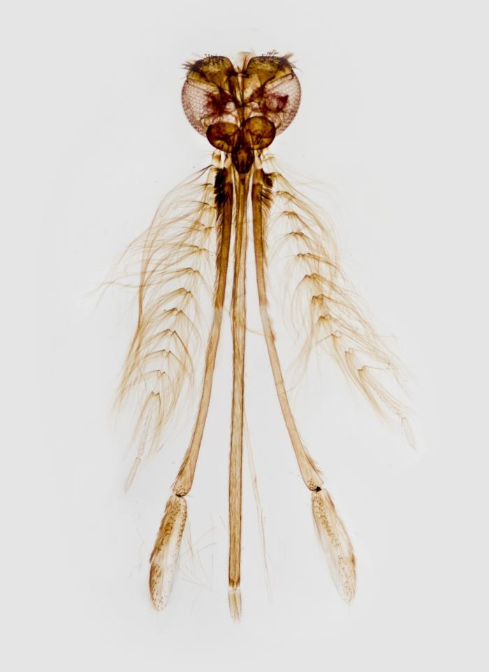
The genus name Anopheles is the Greek word for “harmful” and “useless.” This one genus of mosquitoes has caused incalculable human misery. It can transmit some viruses, but it is infamous for being the only mosquito that can transmit malaria. Its geographic range is global, and it is indeed a very hearty little creature, able to withstand a wide range of temperatures. There are over 450 species of Anopheles; about 30 of them can transmit malaria to humans.
The recorded history of malaria goes back thousands of years, with writings from India, China, Mesopotamia, and Greece showing a strong likelihood of a substantial presence of malaria in many cultures worldwide. Some historians make a solid case that the introduction of malaria into Rome from Africa was a major factor in the Empire's downfall. Many other tales relate to how malaria quite possibly altered history.
The name malaria comes from the Italian words mala, or “bad,” and aria, or “air.” Other words to describe it were ague (acute fever), miasma (pollution or defilement), and paludism (swamp or marsh). The disease most probably originated in Africa in non-human primates then spread to humans and accompanied them as they spread around the world, taking the mosquitoes with them. It most likely spread to the Americas with the opening of trade, particularly the slave trade. The mosquitoes could easily have traveled to the New World in the water containers on the ships and the parasite in the bloodstreams of the native Africans and crews.
The 1880s and 90s were a time of great advancements in understanding the causes of infectious diseases. Pathogens were being discovered at a rapid rate, and information was shared liberally between scientists. But the cause of one of the most terrible diseases in the world escaped discovery. At the time, severe diseases were found to be caused by bacteria: cholera, typhoid, diphtheria, etc. It was assumed that a disease like malaria, with profound chills, fever, and rigors, would also be caused by bacteria. Parasites were not known to cause acute febrile illness. The ones recognized were the helminths, the large worms. The thought that an animal parasite could cause a disease like malaria didn’t seem at all possible. So all attention was given by the top scientists of the day to discovering the bacterial cause of malaria.
One worker took a different approach. Charles Louis Alphonse Laveran was a physician in the French military. His father and grandfather were both men of medicine, and his mother’s father and grandfather were prominent in the French army. After serving in the Franco-Prussian war, Dr. Laveran was posted in Algeria, where he could first-hand examine the blood of a nearly unlimited number of malaria patients. Though his microscope was crude and able to magnify to only around 400x, he described in some detail the presence of tiny crescent-shaped creatures that appeared protozoal, not bacterial. He noted in many a tiny pigment, which we now know to be a breakdown product of hemoglobin that the organism secures from the infected red blood cells.
Dr. Laveran’s description and theory of malaria’s cause were initially met with great skepticism, but other scientists confirmed his findings. Of great help was the use of aniline dyes, especially those developed by Dimitri Leonidovitch Romanowsky in the early 1890s. Romanowsky’s stains were particularly effective in showing the components of blood. We still use the Wright and Giemsa stains, developed from his original. In addition to blood cells, they also show in great detail all the stages of the malarial parasite. Meanwhile, the Carl Zeiss Company developed a high-powered lens that, using a clear oil to reduce refraction of the image, could magnify 1,000 times, giving a much better look at the tiny microbes. With Laveran’s initial discovery and the confirmation achieved by better staining and microscopic technique, it was firmly established that malaria was indeed caused by an animal, albeit a tiny one, known as a protozoon.
The means of acquisition of the disease was as perplexing as its cause. The two obvious ways of getting an infection are food and water or inhalation. But all efforts to demonstrate these means of transmission met with failure. Mosquitoes had been associated with malaria for hundreds of years, but just how was mere speculation. Some thought it might be drinking water in which mosquitoes had died. Others offered the idea that the wind carried dead dried-up organisms to people in the area who breathed them in. Of course, no credible evidence was ever presented for these or other theories.
One theory was that the mosquito wasn’t just a passive carrier of the organism but a vital part of the malarial organism’s life cycle. A prestigious investigator of tropical medicine working in England, Patrick Manson, became intrigued by that possibility and, along with some Italian workers, set about the task of discovering this possibility. While carrying on his medical practice in London, Manson engaged a British Army physician named Ronald Ross to carry out the investigation in the field. Ross was stationed in India and began the work in 1895.
Ross was a military officer first, a malarial researcher second, and the two careers often conflicted. He was also given to writing a novel and some poetry, which didn’t help the malaria investigative cause. His early work involved looking at mosquitoes such as Culex and Aedes, which don’t spread malaria. So after dissecting over a thousand mosquitoes over two years, nothing was learned, and the effort seemed fruitless. But one day, he happened to encounter an Anopheles mosquito which did indeed have evidence of malarial parasites in its abdomen. Just like that, the first inkling of the spread of the disease became known. Through diligent work, he and other workers, mostly the Italians, showed conclusively that Anopheline mosquitoes not only transmitted the disease from person to person but were critical to the pathogen’s reproduction and infectivity.
Laveran and Ross were awarded the Nobel Prize in Physiology or Medicine for their work.
The third very important part of the natural course of the disease to be discovered is what happens after the organism enters the body following a mosquito bite. It was known that the organism travels around the body in the bloodstream, making the patient ill while it multiplies in the red blood cells, then is picked up by another mosquito in which it undergoes sexual reproduction. The cycle is begun anew with another bite. But the incubation time following the bite is around two weeks. What happens to the creature during that time, after the invasion, and before it starts invading the red blood cells? The question eluded researchers for decades.
Unfortunately, a grave mistake was made early in investigating the organism’s pathogenicity, and the wrong course was pursued for many years. A very influential German scientist, Fritz Schaudinn, described and published in 1903 what he claimed to be a direct observation of the malarial sporozoites, the infective units spit out by the mosquito, directly invading red blood cells in the bloodstream. This was held as truth for forty years until 1948, when scientists working at, appropriately, the Ross Institute for Tropical Medicine in London, showed conclusively that the organisms invaded the liver after inoculation and developed there for two weeks before proceeding to enter the bloodstream to infect red blood cells.
Today, students reviewing the life cycle of malarial parasites are given a very concise informative diagram of the events, usually in circular form. Little do many realize the painstaking, often dangerous, work that went into discovering these relatively simple facts. It took over a half-century by some of the world's great scientific minds. It’s not as simple as it looks.
The name assigned to the malarial parasite of humans is Plasmodium, which derives from the Greek words plasma and ode, which mean “molded” or “formed,” and “like.” There are five recognized species of humans, with two predominating, Plasmodium vivax and Plasmodium falciparum. Two other less common species are P. malariae and P. ovale, and a fifth rare species of P. knowlesi. All have the same general life cycle, which is carried out in three stages:
Mating and growth in the mosquito abdomen
Development in the cells of the human liver
Development in the bloodstream
The mosquito provides the parasite double service. For one, they suck blood from the victim and bring it into their bodies along with the malarial parasites. In the mosquito abdomen, the sexual forms of the parasite, the male and female gametes, unite, giving the embryonic form of new organisms. These take a few weeks to develop. They then proceed to the mosquito’s salivary glands. The embryonic organisms in the mosquito are known as an ookinete, which comes from Greek words meaning “mobile egg.” (The word ookinete is an odd one, with about a half dozen possible pronunciations. Two are accepted, o uh kin EET, and o uh KINE eet). The ookinete is mobile by a flagellum, and it works its way up the mosquito's gut as it matures, the embryo growing inside it. When it reaches the mid-gut, the ookinete turns into a cyst. Again, the creatures inside are growing, and when the cyst is ripe, they burst out and “swim” to the mosquito’s salivary gland.
These slithering little creatures are known as sporozoites. The spore part comes from the Greek word meaning “seed,” and the “zoite” suffix indicates that they are indeed animals. While considered spores, they sure don’t look or behave like fungus or bacterial spores that come to mind. They emerge from a cyst, and like a typical spore, they seek a place to implant and begin growing, but they are actively motile and are powered by a dynamic energy system. With many of them in the mosquito salivary gland, they are ready for the next part of the journey.
The sporozoites look like tiny worms, and they have the means of a rather forceful movement, described as gliding motility. The number of sporozoites introduced into the skin by the mosquito varies, anywhere from a dozen to over a hundred. When taking a blood meal, the mosquito must first inject an anticoagulant present in its saliva, and it is with this injection act the sporozoites are pushed out.
When injected by the mosquito into the skin of the unfortunate human victim, the sporozoites begin a downward sojourn until they reach a blood vessel. Some will encounter a macrophage in the skin and be eliminated; some will enter the lymphatic system and make their way to a lymph node, also most likely to be eliminated. But those that reach the blood will eventually find their way to the liver.
The liver is the body's only organ in which sporozoites can carry out their early mission. To ensure they get there, they have a protein that can detect that they are in the right place. They even can travel right through the Kupffer cells, the resident macrophages of the liver, to get to the liver cells, known as hepatocytes. The sporozoites have on their surface a protein (circumsporozoite protein, CSP) that allows them to attach to a receptor on a liver cell that is the same one that receives high-density lipoprotein-bearing cholesterol. Once attached, they penetrate the cell. At that point, the infection has taken root.
When inside the liver cell, the sporozoite is encased in a parasitophorous vacuole. Inside this membrane, it is free to begin reproduction. It also emits proteins that prevent the host cell from undergoing apoptosis, thus maintaining its source of sustenance. The type of malarial reproduction in the liver cell is binary fission, as one cell splits into another, which in turn continues dividing. It’s similar to the replication of bacteria. The number of malarial cells produced in the single liver cell inside the vacuole is impressive, over 30,000. Because the splitting of a single one is the means of making this many daughter cells, the term schizont is applied. The Greek word for “split” or “divide” is skhizein. It is often used in English as the prefix schizo-. Each tiny member of the enormous schizont is given the term merozoite, the “mero,” taken from the Greek word meros, meaning part or share. The same terms are applied to the parasite when they are in the bloodstream.
At a certain point, the burden of these rapidly growing merozoites overwhelms the liver cell. One side of it opens, and a vast number of merozoites come pouring out into the bloodstream. They make it to the lungs. There, a capsule surrounding the cell is removed, and they are freed into the bloodstream. From there, entering a red blood cell is a simple matter.
Malarial merozoites have a front and back end. On the front end, they have attachment and red cell penetration devices that easily allow them to enter the red blood cell. Once inside, they begin to gather nutrients from hemoglobin. At this stage, the merozoite has become a trophozoite, the actively feeding stage. Trophozoite comes from the Greek word trophe, meaning “nourishment.” After it has been well fed, the trophozoite begins the division process anew, forming another schizont, this time in the red blood cell. The number of merozoites making up the red blood cell schizont is nowhere near the number seen in liver cells, anywhere from 6 to 36, depending on the species. (Counting the number of merozoites is very useful in the laboratory for identifying the malarial species present). Just like in the liver, each little organism is termed a merozoite. At maturation, the red blood cell bursts open, and the merozoites come pouring out, each capable of infecting another red blood cell.
The merozoites do not spend much time exposed to the circulatory system. It takes less than a minute for one to enter a red blood cell, and there is no limit to the number of cells that may be infected.
This cycle keeps getting repeated. The tiny merozoites quickly adhere to and enter red blood cells, and the red blood cells get ripped to shreds as the dozen or more newly created merozoites exit. Depending on the malarial species, it takes a few days for the red blood cell growth phase to progress.
The symptoms of malaria are mainly due to the inflammatory cytokines, especially tumor necrosis factor-a (TNF-a). The induction of chills, fevers, and rigors is primarily the result of the monocytes of the blood encountering fragmented red blood cells as well as the occasional merozoite and releasing potent inflammatory cytokines.
The bodily organ tasked with removing defective red blood cells is the spleen. The spleen becomes greatly overworked with the tremendous number of defective red cells, often resulting in marked swelling. An enlarged spleen, known as splenomegaly, is one of the hallmarks of malaria.
This cycle of malarial merozoites being released, re-infecting, then again being released goes on for some time, often a couple of weeks. For some reason that is not understood, some of the merozoites do not re-infect another red blood cell after their release but instead mature into either a male or female gametocyte. When a mosquito comes along and takes a blood meal from an active malaria patient, the merozoites it picks up are irrelevant. For the parasite, the only things that matter at this point are the gametocytes that will go on to come together in the mosquito’s gut to produce the ookinete, potentially leading to another person becoming infected.
It’s no wonder it took about 50 years to discover this elaborate life cycle of Plasmodium. So many invasive steps, organism transformations, maturation, and transmission cycles that one gets mixed up just thinking about it. But the entire process has been going on successfully for millennia and is just as vigorous today as it was in ancient times.
A major obstacle to any invading microorganism is the host’s immune response. The human immune system is amply equipped to combat invading organisms with weapons such as complement and other serum proteins, phagocytic cells, cytotoxic lymphocytes, antibodies, and apoptosis. Clearly, though, the Plasmodium causing malaria gets through, often in no small way. Massive numbers of parasites circulate in the body when the disease is at its apex, sometimes taking weeks for the infection to clear. Sometimes the disease progresses almost unnoticed, with few symptoms, while the disease is profoundly debilitating to many. Some patients, especially pregnant women and children, die.
One of the organism’s advantages is its quickness. They don’t stay exposed in the tissues or bloodstream for long. Being intracellular pathogens (they reproduce inside a living cell), they are protected in their habitat in two ways. One is in the liver, where they form a parasitophorous vacuole (PV), a membrane surrounding them, while they form a schizont. The PV cannot unite with the liver cell’s lysosome, so the parasites can develop inside the cell with impunity. Plasmodium also prevents the host liver cell from killing itself through apoptosis, but other cellular functions are carried on near-normal, not to alert any natural killer lymphocytes.
The cell for their asexual reproduction is the circulating red blood cell. These cells lack nuclei, so there is no signaling to the genome to release cytokines or other signaling molecules. RBCs are not attacked by natural killer lymphocytes or cytotoxic CD8 or T-helper CD4 lymphocytes. Damaged RBCs are removed from the circulation, mainly in the spleen and liver, but the parasites have found a way around that.
When a red blood cell floats through the spleen's red pulp, it must be skinny and pliable enough to pass through. The spleen effectively removes, disables, and recovers components of RBCs that don’t measure up. On a routine basis, this usually means RBCs that are too old. It’s a common, necessary procedure. A red blood cell infected by Plasmodium is not likely to safely make it through the spleen, so one species, falciparum, the deadliest malarial parasite, has a solution: don’t circulate through the spleen. P. falciparum manufactures at least three proteins expressed on the surface of the red blood cell it is infecting. They cause the RBC to adhere to various surfaces, including the endothelial lining of small blood vessels, other red blood cells, and some types of lymphocytes.
By attaching to the lining of venules, the organism inside its host red blood cell is not swept along with the circulation. It just stays in one place as it matures, forming its schizont. Other red blood cells are infected when the schizont bursts and the merozoites are released. They circulate, but some make it through the spleen because the red cell deformity is not advanced in newly infected cells. This adhesion is the reason for the peculiar, characteristic fact about falciparum malaria: in the peripheral blood, only the early ring forms (single merozoite) stage is seen, not the more mature trophozoite and schizont stage.
Other red blood cells are another surface the falciparum adhesion molecules can stick to. The result is a clump of RBCs known as a rosette. The formation of a rosette serves two purposes for the parasite: the cluster will get stuck in the small venules, preventing circulation through the spleen, and it also provides protection from complement and phagocytes attacking the infected cell. Having all these normal but stuck RBCs around it shields the parasite’s surface proteins from recognition by members of the immune system.
Malarial parasites, of course, depend on mosquitoes to assist them in propagating the species. Sometimes the mosquitoes aren’t there, either because of the weather or another reason. The adherence proteins may help the parasite “hide out” in the host for a prolonged period and produce gametocytes at a low level for a while, at least until the mosquitoes return.
Plasmodium feeds on the hemoglobin of the red blood cell that it invades. The hemoglobin is broken down into two major components: globulin, the protein portion, and heme, the iron-containing section. The protein portion is digested by the parasite into its component amino acids. The heme portion presents the parasite a problem. Plasmodium doesn’t use all of it, so the excess is freed during the infection. It is toxic and must be taken care of. The organism has the means to aggregate the free hemoglobin into insoluble crystals, called hemozoin, which can float free to be picked up and metabolized by monocytes. This is beneficial to both the parasite as well as the host. In some heavy infections, however, especially those caused by Plasmodium falciparum, the release of heme is not controlled, and there is considerable tissue damage to the host, particularly the kidney. The result is a condition known as “blackwater fever,” a lurid description of the color of the urine. Often, this condition is fatal.
The serum aggregate of proteins known as complement is an important deterrent to many potential invading organisms, including Plasmodium. It is a potent weapon, and when unleashed, it can destroy any type of cell, including our own. To prevent self-destruction by the highly detrimental complement component C3b, we are equipped with an influential protein known as Factor H. It is a regulator protein that associates itself with human cell surfaces, rapidly deactivating the destructive portion of complement. Some malarial parasites have created their own version of Factor H, thus rendering themselves much less vulnerable to complement’s destructive powers.
Several chemical compounds have been discovered and modified through the ages to combat malaria. The first was quinine, derived from the bark of the cinchona tree of South America. Quinine had its problems, notably unreliable dosages in the early days and toxicity, but it was a reasonably effective drug for hundreds of years. In the 1930s and 40s great effort was made to find an alternative to quinine, or at least modify it to give it a more standard dose and reduce its toxicity.
Quite a few drugs were developed, most of them variations on the chemical structure of quinine. The one that has survived is chloroquine and its close modification, hydroxychloroquine. They are still used effectively today, particularly for prophylaxis, although drug resistance by Plasmodium has become a significant problem. Heme, the iron-containing part of hemoglobin, is toxic to Plasmodium. That’s why it goes to great lengths to detoxify it by converting it into large hemozoin crystals. Chloroquine, an alkaline compound, enters the food vacuole where heme metabolism occurs, preventing hemozoin formation. The build-up of free heme is toxic to the organism. Drug resistance in the parasite is probably due to the inability of chloroquine to attach to and enter the food vacuole or an efflux pump developed by the organism to excrete it.
Another important anti-malarial drug has a fascinating history, just as quinine does. The sweet woodworm plant Artemisia annui has been used for thousands of years in traditional Chinese medicine. Known in China as Qinghao, it was mentioned as an anti-malarial drug in the Handbook of Prescriptions for Emergency, written during the Eastern Jin Dynasty, 317-420 A.D. During the Vietnam War, malaria was a severe problem for all involved, and chloroquine resistance by the pathogen was becoming commonplace. The Chinese government enlisted Dr. Youyou Tu, an expert in traditional Chinese and Western medicine, to seek alternative therapy. Dr. Tu and her colleagues made a thorough search of over 2000 Chinese herbs as well as a study of ancient writings. They came up with what we now call Artemisinin. After experimenting with extraction techniques, the drug in a relatively pure form showed itself very effective in curing acute malaria patients. Various chemical modifications to the core molecule have yielded several effective drugs, which are prominently used today.
The mode of action of Artemisinin has been hard to pin down, but it appears to resemble that of chloroquine, that is, disruption of the formation of hemozoin by the parasite with the resulting killing of the organism. Unfortunately, as with other anti-malarial drugs, resistance has shown up in several endemic areas.
Malaria due to Plasmodium vivax and P. malariae sometimes manifests with a delayed liver stage. Called hypnozoites, the merozoites do not emerge from the liver cell early in the disease but can exist in a dormant state, free to enter the bloodstream later. The drug primaquine is often included in the regimen when treating either of these two species, as it is effective against hypnozoites.
Malaria has been a scourge of humanity for thousands of years. Despite our extensive knowledge of the life cycle of the parasite and the availability of anti-malarial drugs, it remains a problem for about 40% of the world’s population. Hundreds of millions are sickened yearly, and over half a million, primarily pregnant women and children, die. After World War II, the United States embarked on an ambitious program to eradicate malaria from its shores. Patients with the disease were put in strict isolation to keep them away from mosquitoes and given chloroquine to rid them of the parasite. Extensive mosquito-infested areas were sprayed with DDT to vastly decrease the mosquito population. It worked. Today malaria is not seen in the United States, despite the presence of Anopheline mosquitoes. When the mosquitoes take a blood meal, there are no Plasmodium gametocytes for them to take up. The cycle has been broken. Now, many mosquitoes are resistant to DDT, which in many areas is a banned substance, and many organisms are resistant to chloroquine and artemisinin, so this technique is no longer possible.
Developing a vaccine for any microbe is difficult, but making an effective one for parasites is even more challenging. For malaria, the organism undergoes numerous biological changes, and knowing which stage to target is not easy. In the early 2020s, a vaccine was introduced against Plasmodium falciparum, the species with the highest mortality rate. When in the sporozoite stage, the one that enters the body from the mosquito and enters the liver cells, there is a protein on the parasite, circumsporozoite protein (CSP). It attaches to a receptor on the liver cell enabling the parasite to enter. By adding a portion of CSP to an inactive hepatitis B virus, antibodies are formed to CSP, and the activity of the sporozoite is greatly diminished. An adjuvant was added to boost the immune response. In addition to the B-lymphocyte response creating antibodies to CSP, there is also a vigorous T-cell response, with killer lymphocytes primed to eliminate the liver cells that harbor the invasive sporozoites.
While early vaccine trials have been promising, malarial parasites are notorious for mutating and developing resistance to chemical agents. It remains to be seen if they can show similar resistance to the vaccine. CSP is highly conserved, and if it is to function correctly for the parasite, it may have to remain unchanged. Time will tell.
Anopheles mosquito (PHIL)

